Artificial Airways


Gallery Overview
An overview of oropharyngeal and nasopharyngeal airways, endotracheal tubes, tracheostomy tubes, and related devices.


Oropharyngeal Airways
Oropharyngeal airways are featured in this section.
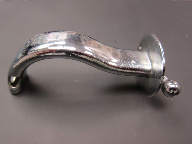
Waters Oropharyngeal Airway
Before plastic was readily available, orophayngeal airways were constructed of metal.
Image from Felix Khusid
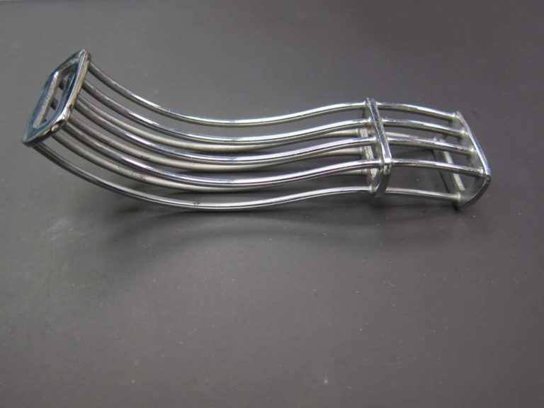
1912 Lumbard Airway
Dr. Joseph Lumbard introduced the "tongue controller" airway in 1912.
Image from Felix Khishid
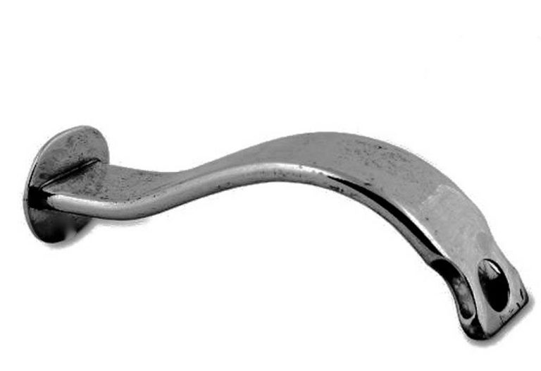
Connell Airway
A metal Connell airway is shown.
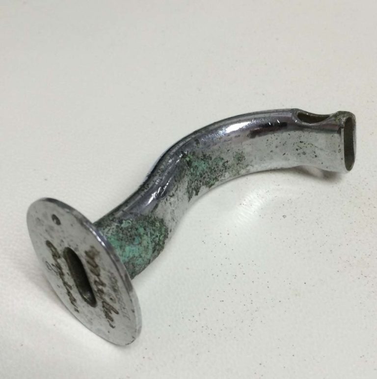
#2 Oral Airway
A metal, #2 oropharyngeal airway is shown.
Image from Gene Gantt
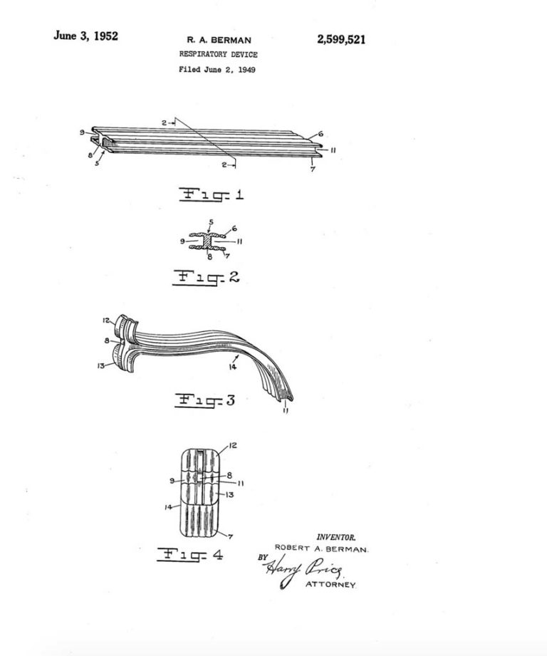
Berman Patent
On June 2, 1949, Robert A. Berman applied for a patent for his respiratory device, a non-metallic oropharyngeal airway. The patent was awarded on June 3, 1952.
Image US Patent and Tradmark Office
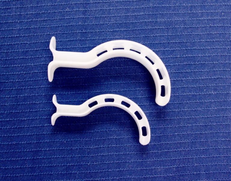
Berman Airways
Berman oropharyngeal airways are shown.
Image from Gayle Carr
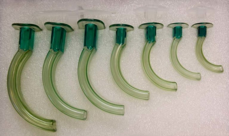
Guedel Airways
Guedel oropharyngeal airways are shown.
Image from Gayle Carr
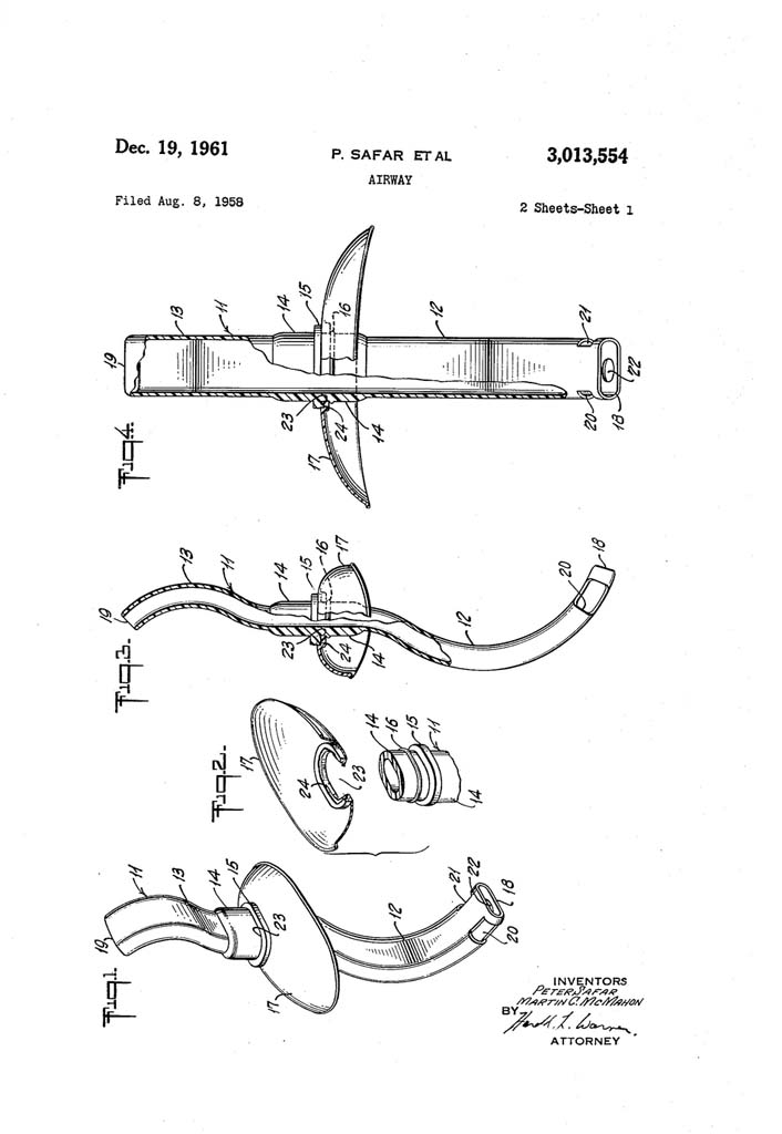
Safar's "S" Airway
On August 8, 1958, Peter Safar and Martin McMahon filed a patent application for "Airway". The patent was granted on December 19, 1961.


Nasopharyngeal Airways
Nasopharyngeal airways are featured in this section.
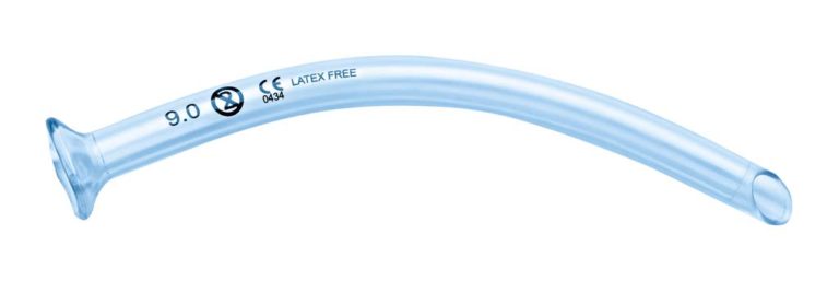

Nasopharyngeal Airway
A nasopharyngeal airway is shown.


Endotracheal Tubes
Endotracheal tubes are featured in this section.

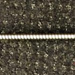
Metal ET Tube
A metal, pediatric endotracheal tube, owned by Felix Khusid, is shown.
Image from Felix Khusid
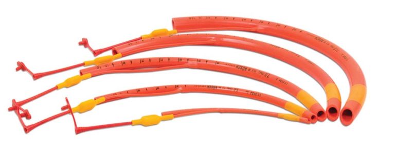
Rusch ET Tubes
Several "red rubber" Rusch endotracheal tubes are shown. The cuffs were low volume, high pressure.
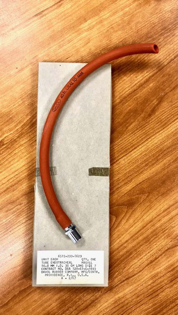
Davol ET Tube
An uncuffed, Davol endotracheal tube , circa 1967, is shown.
Image from Felix Khusid
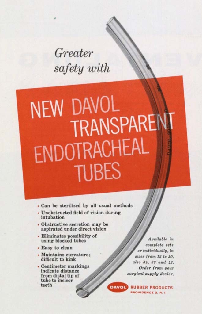

1958 Transparent ET Tube
This ad for transparent endotracheal tubes appeared in the September 1958 issue of the INHALATION THERAPY journal.
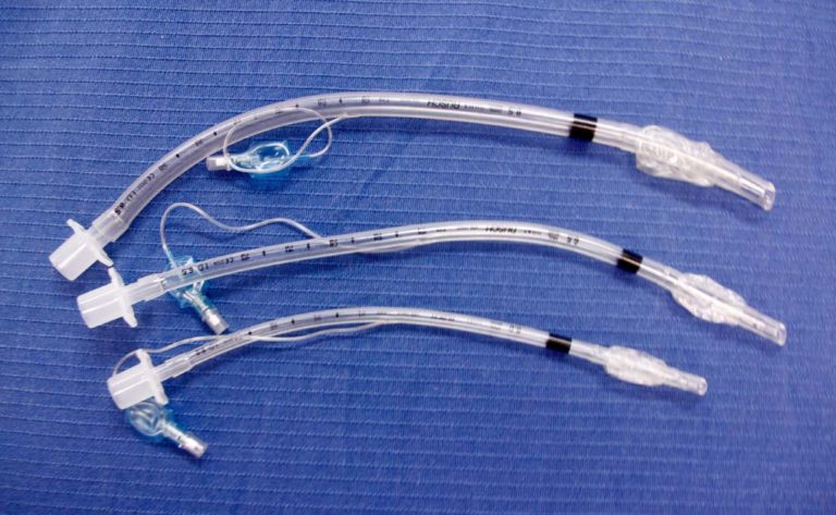
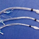
Cuffed ET Tubes
Several sizes of clear, cuffed ET tubes are displayed.
Image from Gayle Carr
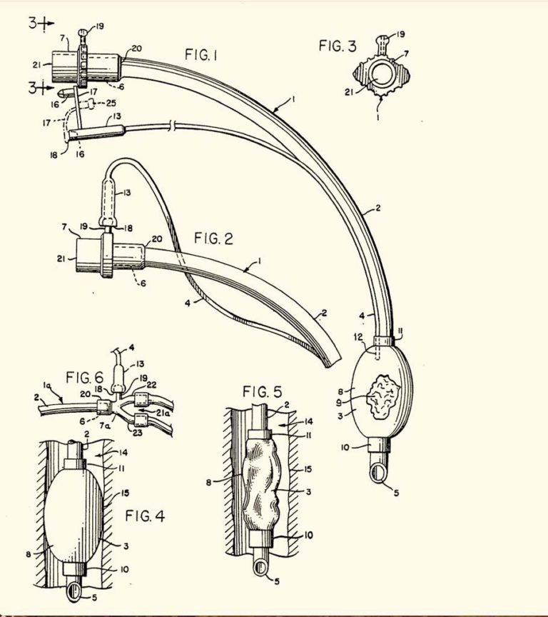
Bivona Cuff
Bivona, Inc. filed filed a patent application on August 9, 1982 for a "Tracheal Tubes" invented by Seymour W. Shapiro. The patent , EP0072230 B1, was granted November 5, 1986.
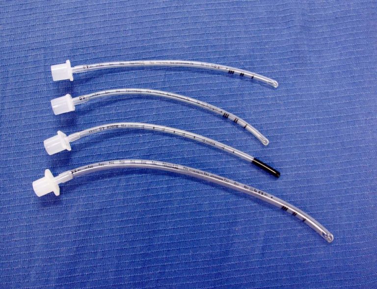
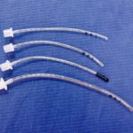
Uncuffed ET Tubes
Several sizes of uncuffed ET tubes are shown.
Image from Gayle Carr
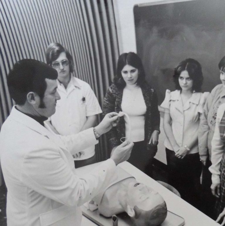
1975 Intubation Lab
Charles McKnight is shown in this 1975 photo preparing his respiratory therapy students for an intubation skills lab at Lutheran Hospital, Moline, Illinois.
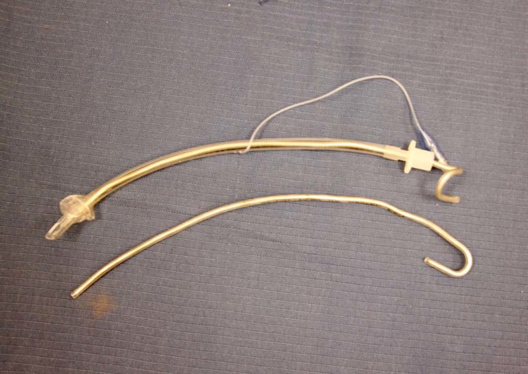
ET Tube and Stylette
A cuffed ET tube and metal stylette is shown.
Image from Illinois Central College Archives, 1999
Duo Cuff ET Tube
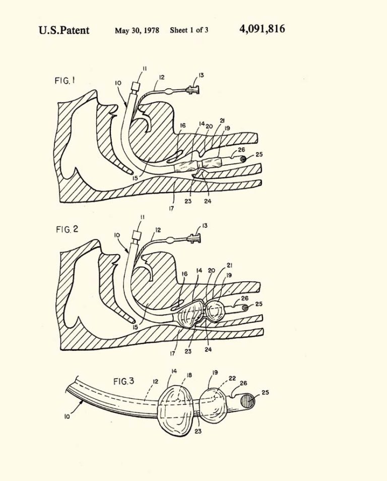
James O. Elam was awarded a patent on May 30, 1978 for his invention of a double cuffed, endotracheal tube.
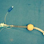
CombiTube
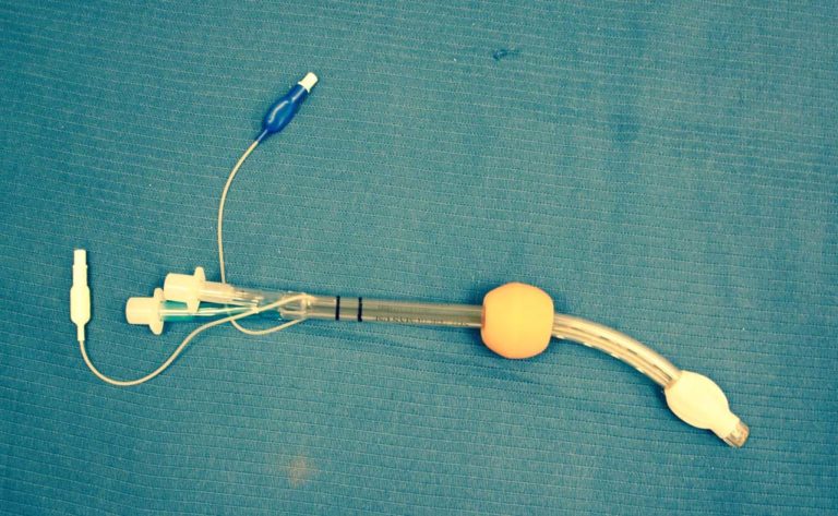
The esophageal tracheal Combitube is shown.
Image from Illinois Central College Archives, 1999
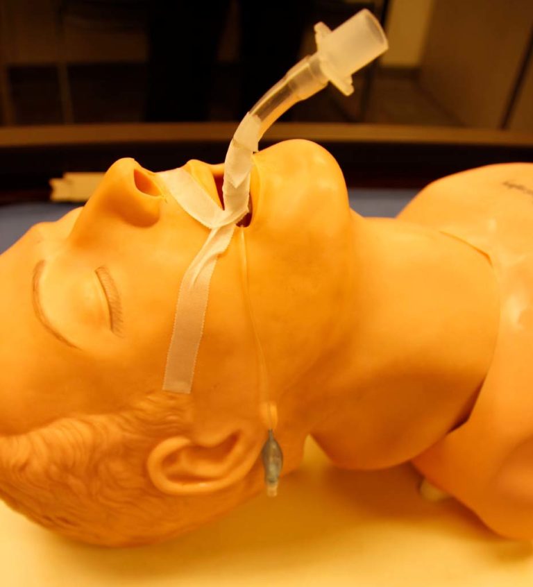
ETT Securement by Tape
One method of securing an ET tube is demonstrated on a manikin.
Image from Illinois Central College Archives, 1999
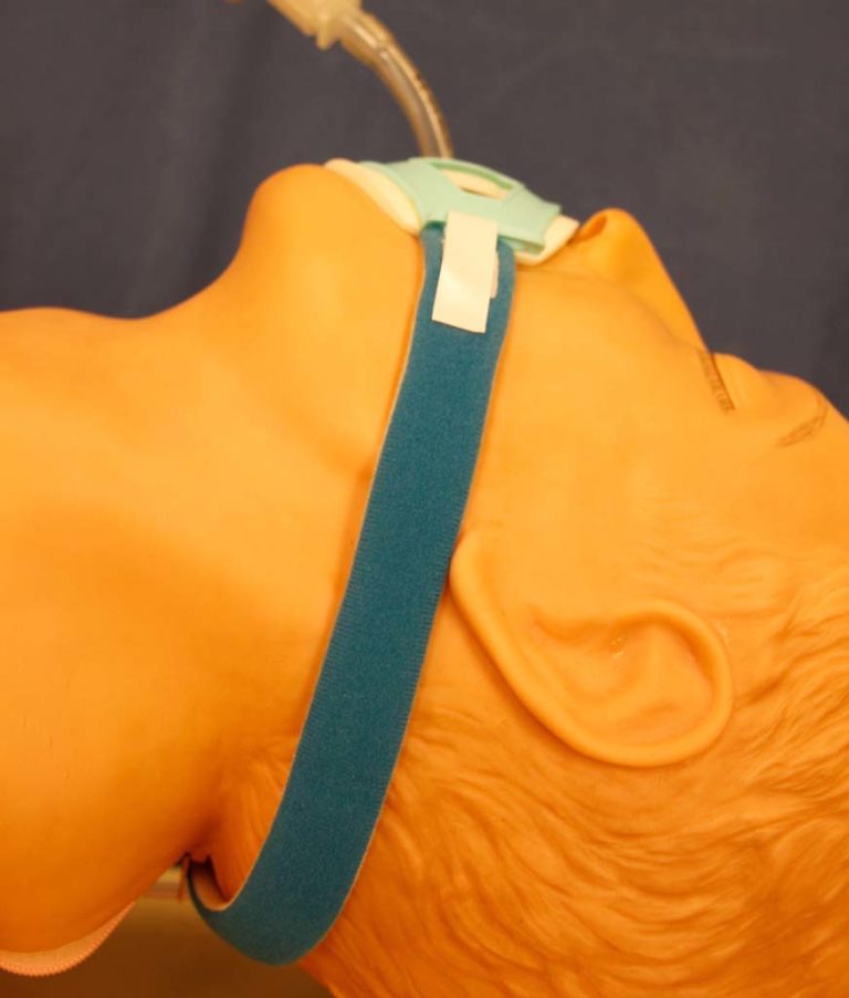
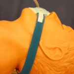
ETT Holder
An adjustable endotracheal tube holder is shown on a manikin.
Image from Illinois Central College Archives, 1999
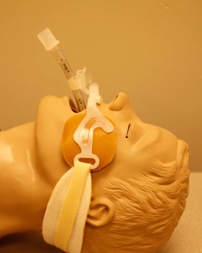
ET Tube Holder
An endotracheal tube holder with cushioned padding is shown on a manikin.


Laryngoscopes
Laryngoscopes are featured in this section.
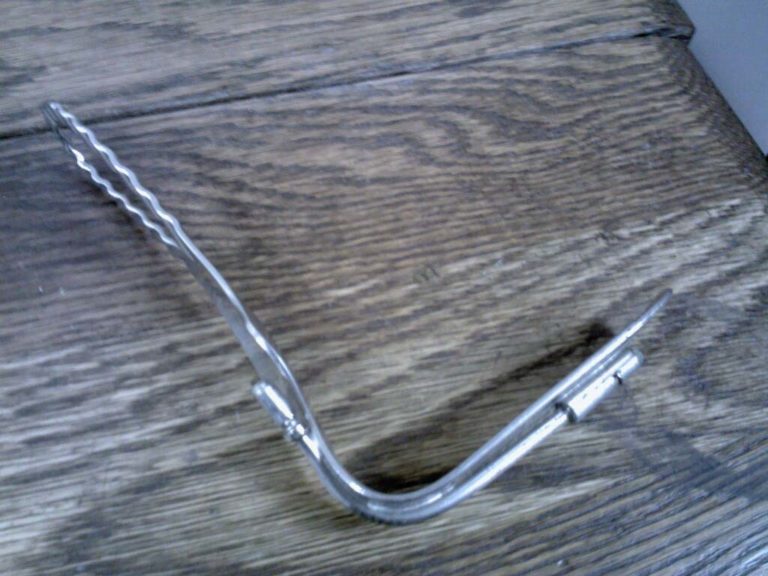
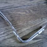
Early Laryngoscope
This early laryngoscope is from the collection of Gene Gantt.
Image from Gene Gantt
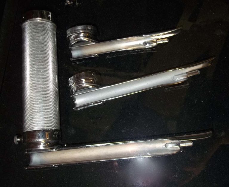
Laryngoscope
A laryngoscope handle and blades are shown.
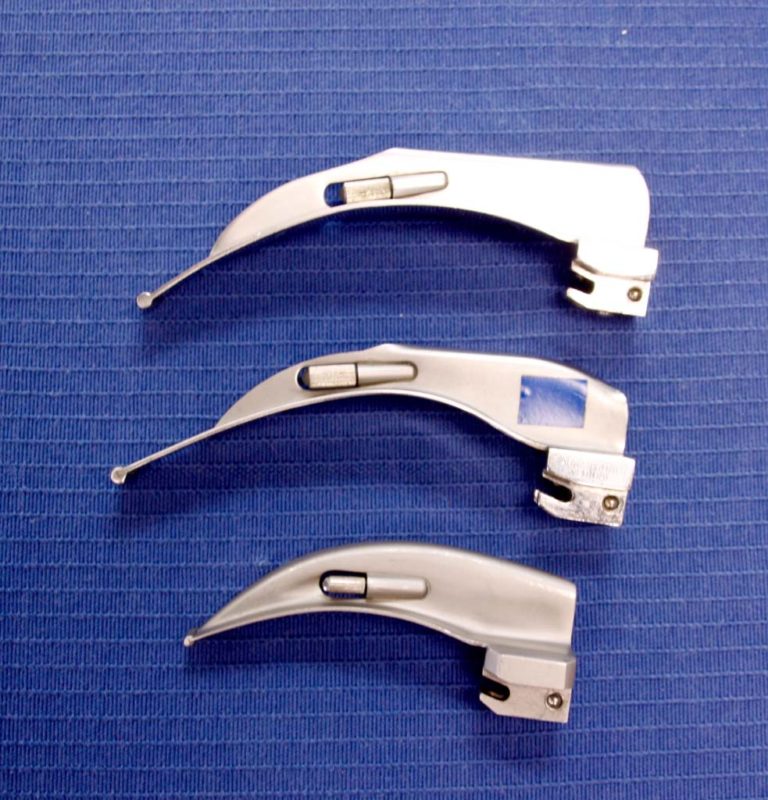
MacIntosh Blades
MacIntosh blades of various sizes are shown.
Image from Gayle Carr
Magill Blades
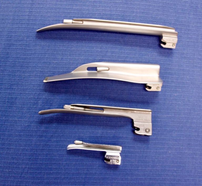
An assortment of sizes of Magill blades are shown.
Image from Gayle Carr
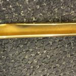
Laryngoscope Blade
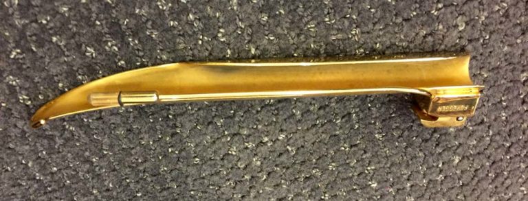
This image of a golden laryngoscope blade was contributed by Felix Khusid.
Image from Felix Khusid


Tracheostomy Tubes
Tracheostomy tubes are featured in this section.
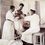
1916 Tracheotomy
This 1916 photo from a collection of historical medical photos was captioned:
"Tracheostomy Operation, Performed by a Russian Military Surgeon Poland. As the Russian surgeon steadies his knife two assistants steady the patient. The doctor is about to perform a tracheostomy with the patient sitting up. During wartime procedures were often performed in unorthodox ways."
"Tracheostomy Operation, Performed by a Russian Military Surgeon Poland. As the Russian surgeon steadies his knife two assistants steady the patient. The doctor is about to perform a tracheostomy with the patient sitting up. During wartime procedures were often performed in unorthodox ways."
Image from a collection shared by Aracely Bigelow
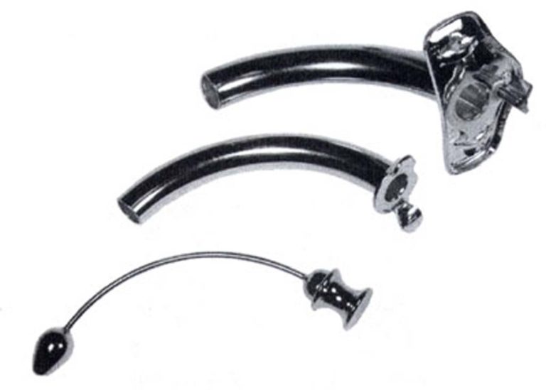
Uncuffed Jackson Trach Tube
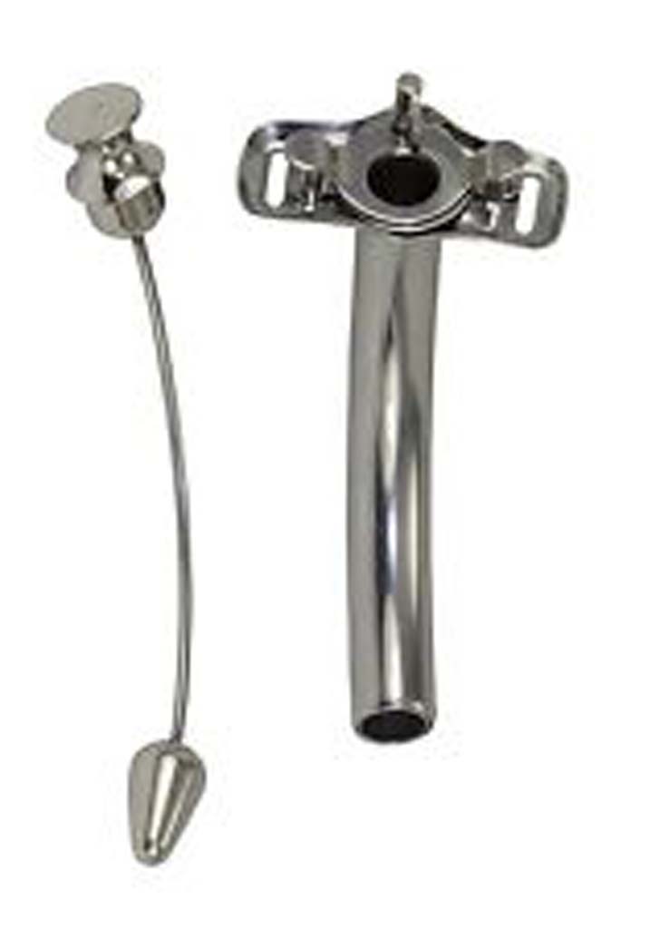
The components of an uncuffed tube are shown: outer cannula, inner cannula, and obturator.
Jackson Uncuffed Trach Tube
A Jackson uncuffed trach tube and obturator are shown.
Image from Gayle Carr
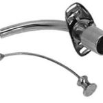
Trach tube with 15 mm adapter
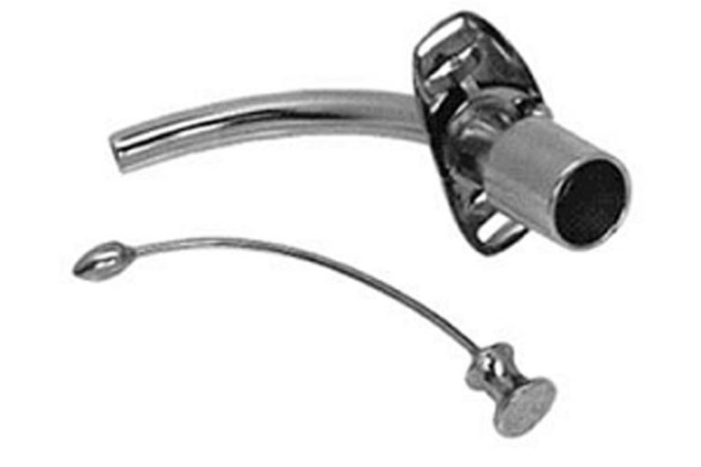
A metal trach tube with a 15 mm adapter attached to the inner cannula is shown. The obturator is also pictured.
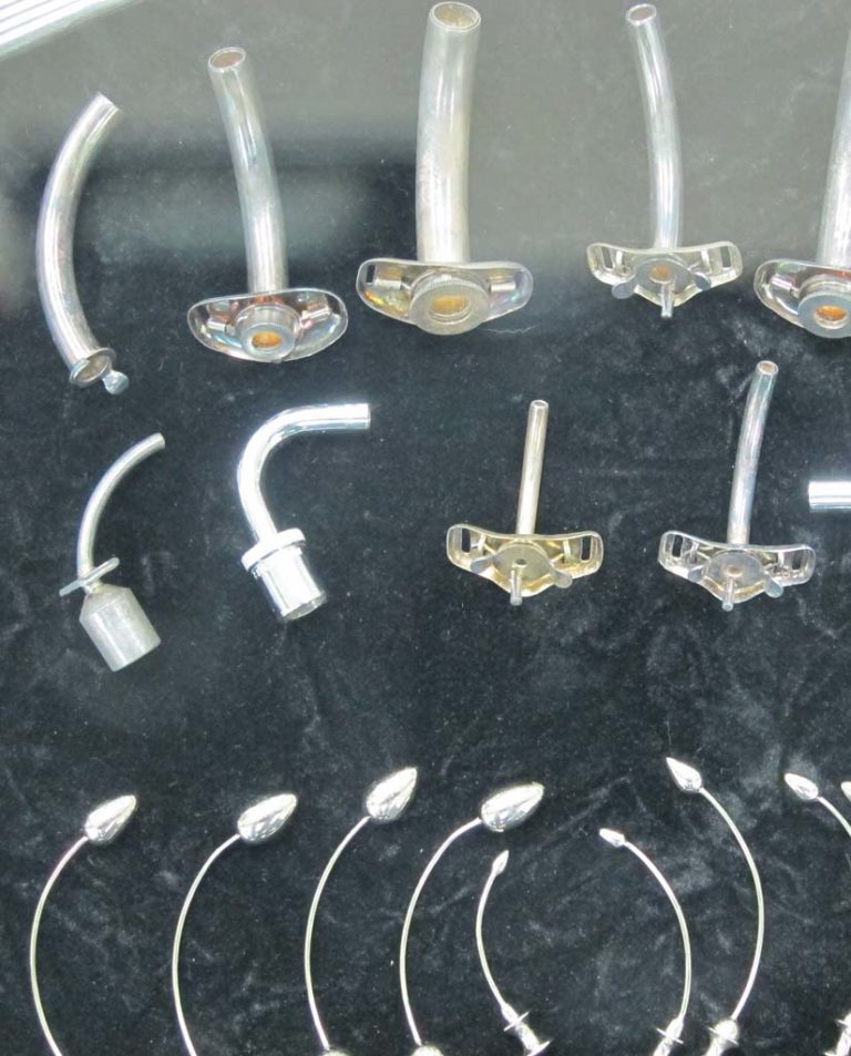
Trach Tubes
An assortment of trach tubes are shown.
Image from Felix Khusid
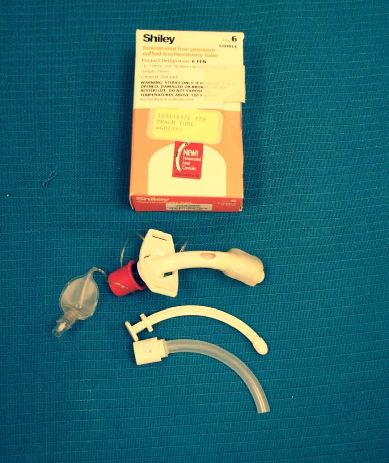
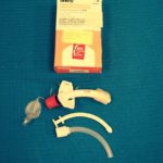
Fenestrated Trach Tube
A Shiley fenestrated, cuffed trach tube is shown.
Image from Illinois Central College Archives, 1999
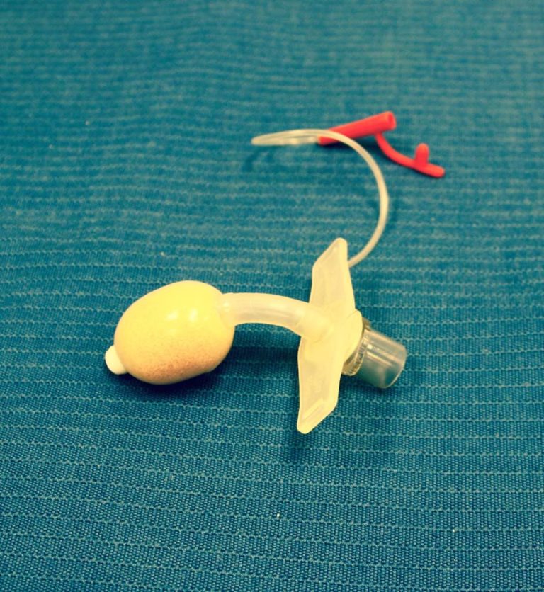
Bivona Fome Cuff
A Bivona fome-cuff trach tube is shown.
Image from Illinois Central College Archives, 1999
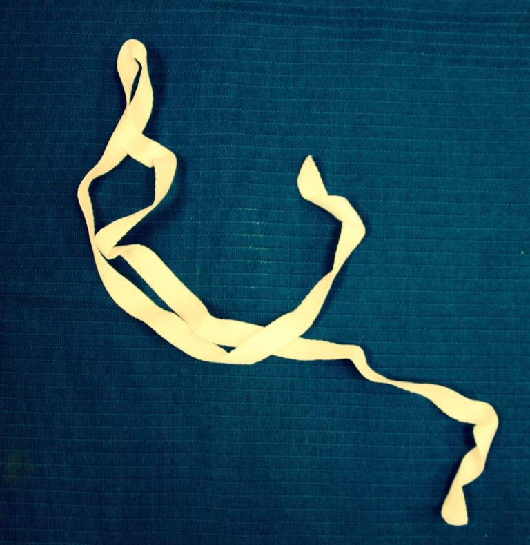
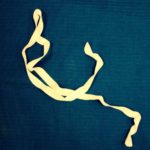
Twill Ties
Cotton twill tape can be used to secure the outer cannula of a trach tube.
Image from Illinois Central College Archives, 1999
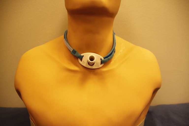
Trach Tube Holder
An adjustable, tracheostomy tube holder is shown.
Image from Illinois Central College Archives, 1999
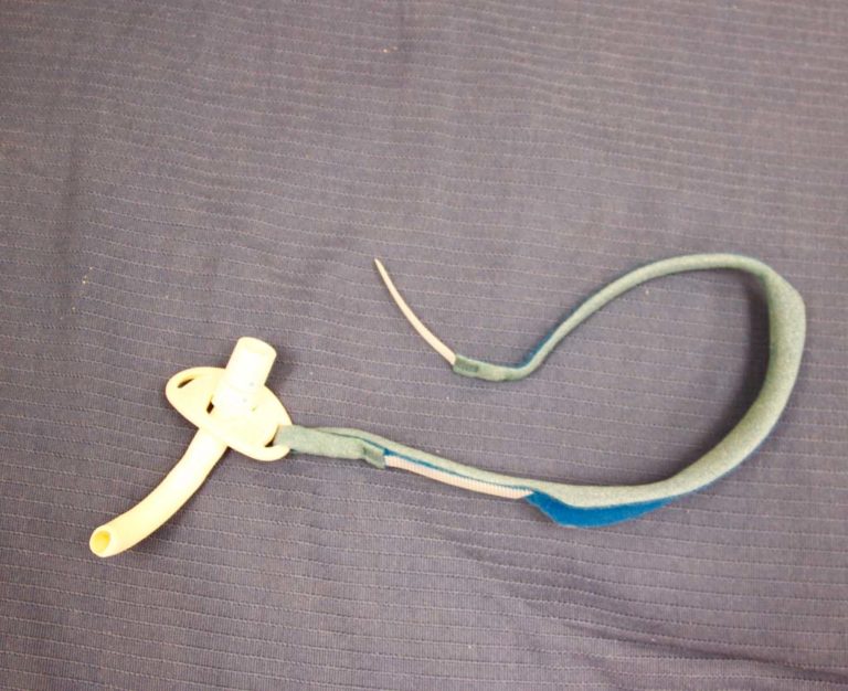
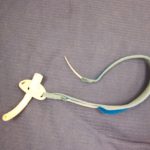
Trach Tube Holder
An adjustable, padded trach tube holder is shown.
Image from Illinois Central College Archives, 1999


Auxiliary Devices
Auxiliary devices for artificial airway management are featured.
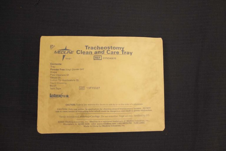
Trach Care Kit
The components of a cleaning kit for trach care are listed on the front of this kit.
Image from Illinois Central College Archives, 1999
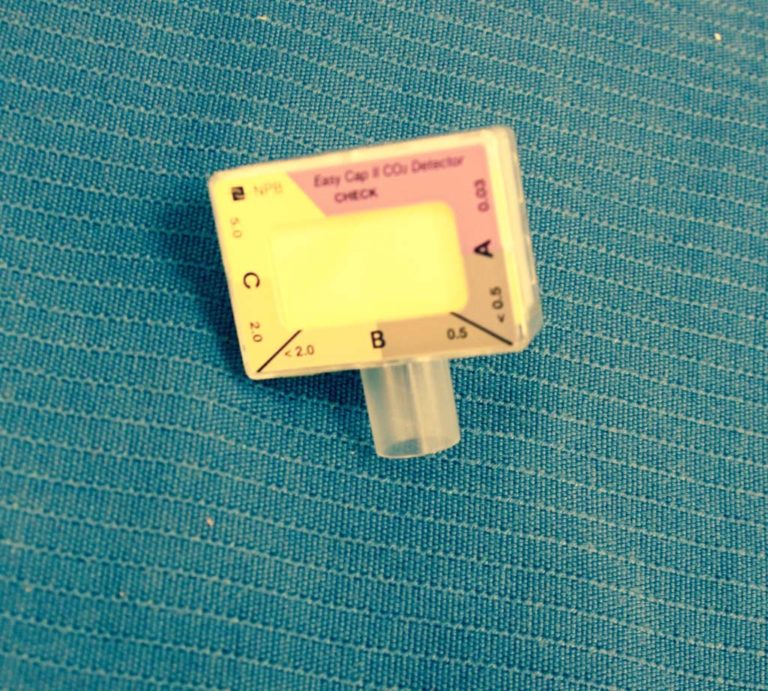
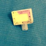
Easy Cap CO2 Detector
A colorimetric CO2 detector is used to confirm ET tube placement. The device changes color when CO2 is detected from the ET tube.
Image from Illinois Central College Archives, 1999
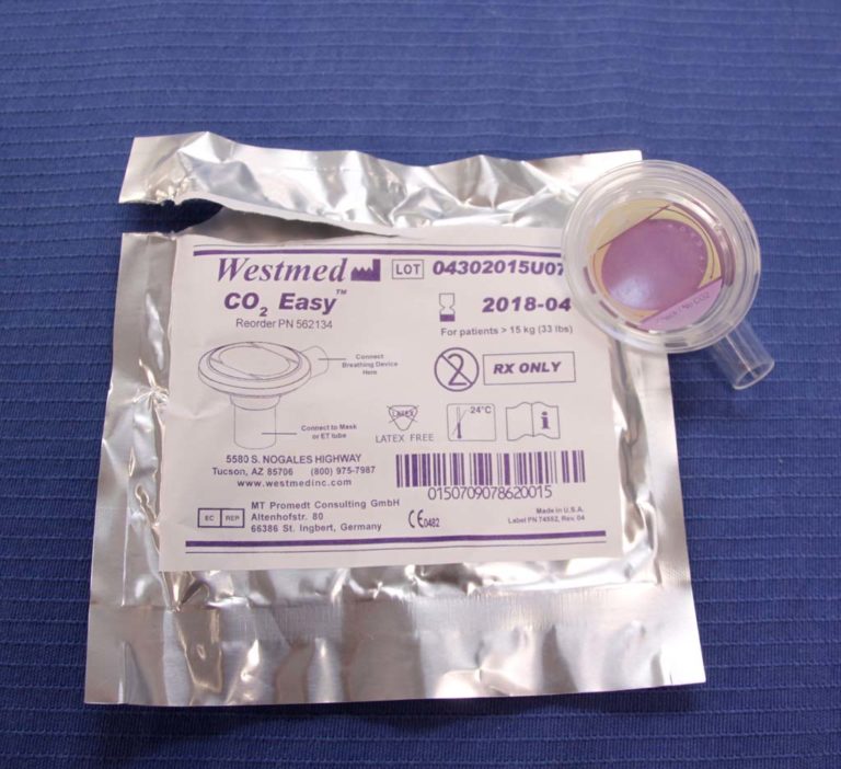
CO2 Indicator
A colorimetric CO2 indicator is utilized to confirm that an endotracheal tube is positioned in the airway and not the esophagus.
Image from Gayle Carr
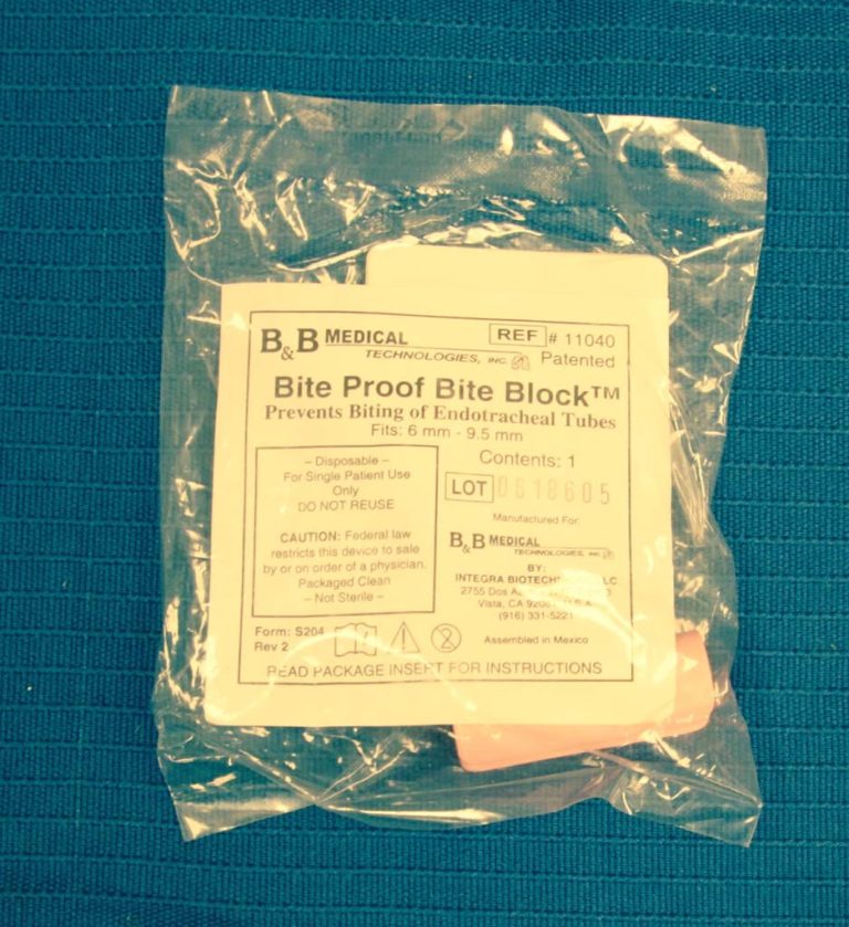

Bite Block
A bite block can be inserted to prevent a patient from biting an oral endotracheal tube.
Image from Illinois Central College Archives, 1999
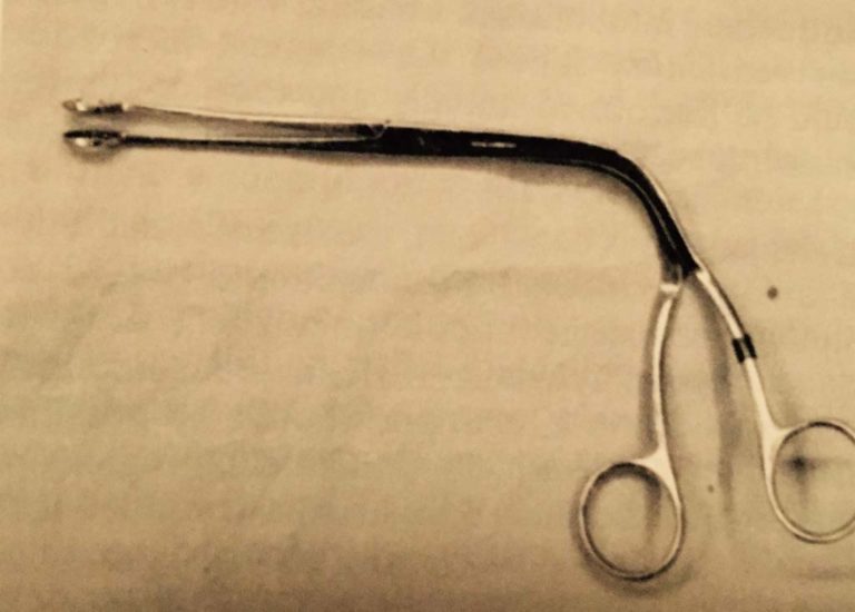
Magill Forceps
A Magill forceps may be required to assist with the placement of a nasoendotracheal tube.
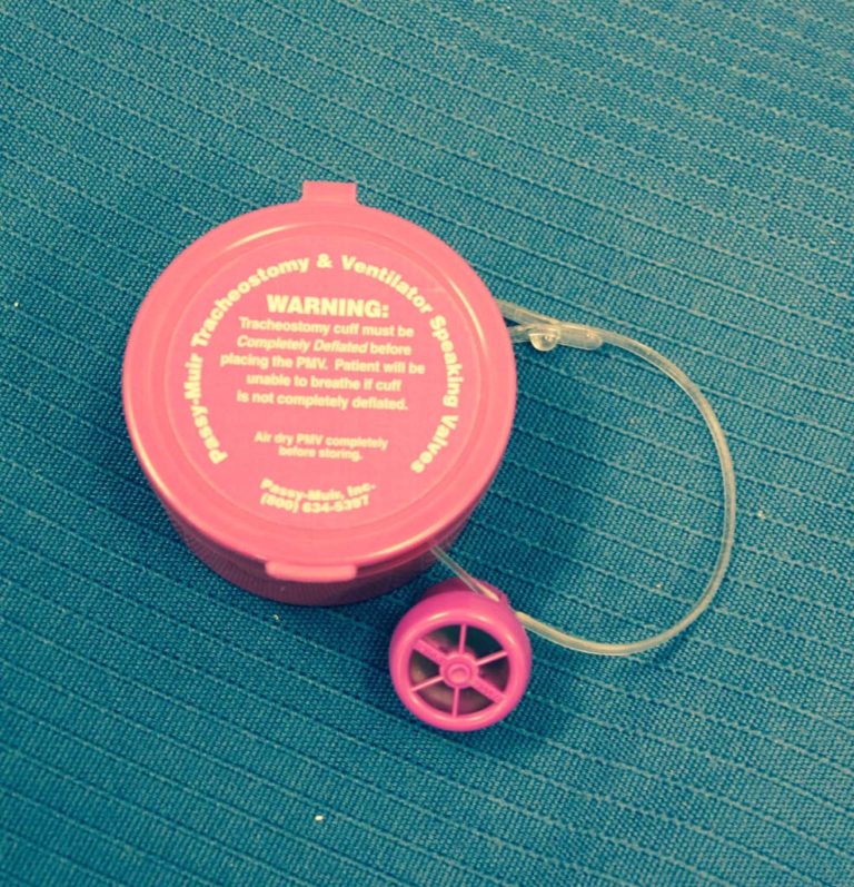
Passy-Muir Speaking Valve
The Passy Muir Valves are used to facilitate speech in patients in patients with tracheostomies and in ventilator dependent patients. The device was designed by David Muir in the mid-1980s after he spend several months on a ventilator and was frustrated by the inability to speak.
Image from Illinois Central College Archives, 1999
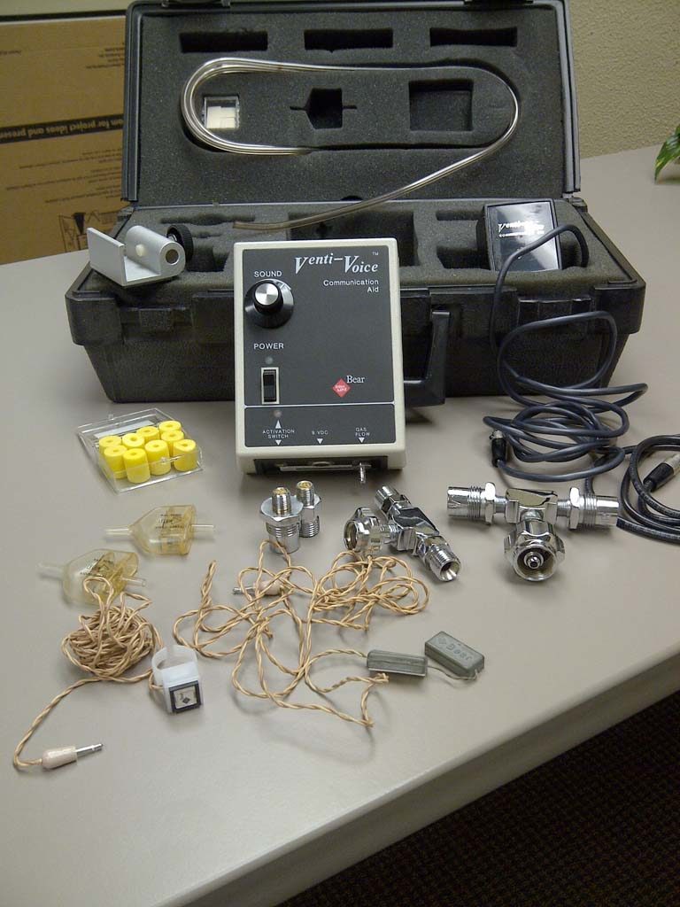
Venti-Voice
BEAR Medical's Venti-Voice Communication Aid circa 1980 is shown. Venti-Voice was designed as a speaking aid for people dependent upon a ventilator but could be operated independent of a ventilator. The user activated a flow of gas with a hand-held switch or a magnetic forehead switch which allowed gas flow to a tone generator.
Image from Cheryl Hoerr
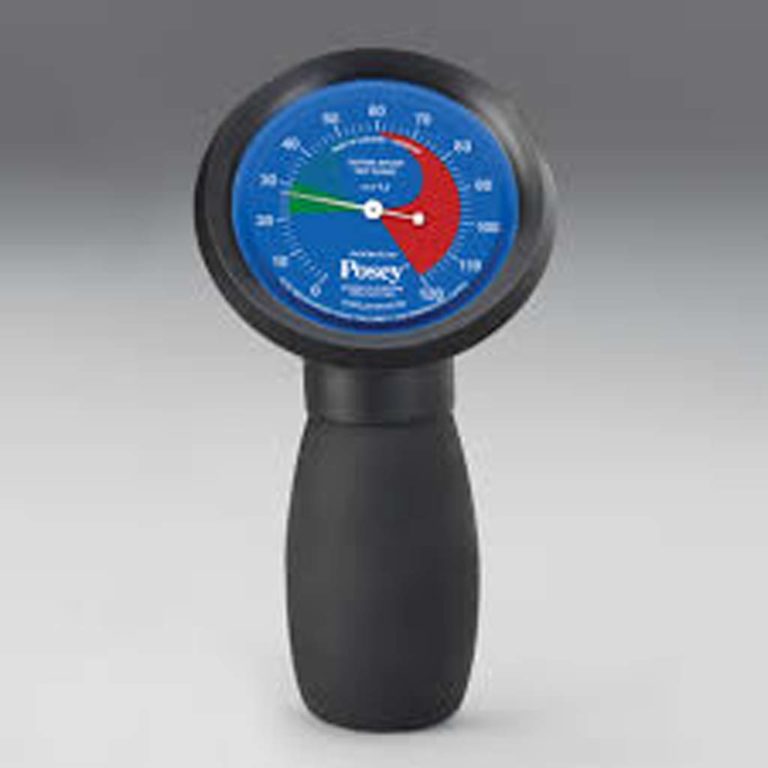
Posey Cufflator
The Posey Cufflator could be used to inflate or deflate endotracheal cuffs as well as monitor the intracuff pressure.

Images to share?
Please contact us if you have images to contribute to this gallery.